
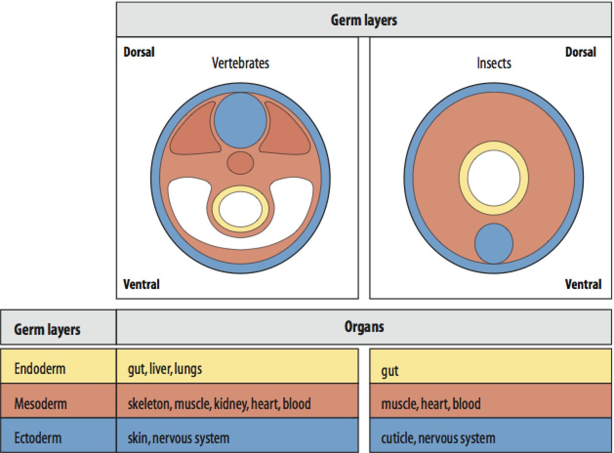
Gastrulation process of animal embryogenesis produces cellular rearrangements giving rise to three primary germ layers called ectoderm, mesoderm and endoderm. The ectoderm, the outermost layer is the first layer to appear.
The lower phyla like Porifera , Ctenophora ,Cnidaria contain two primary layers that give rise to all the tissues and organs of the animals making them diploblastic. The higher phyla groups produce a third layer mesoderm in between making them triploblastic.
The ectoderm in vertebrates basically consists of three parts- the external ectoderm or surface ectoderm, the neural crest, and the neural tube; the latter two are together referred as neuroectoderm.
The neural crest in between contains tissue that will become neurons and glial cells of the adult autonomic nervous system. The neural tube is the precursor to the adult central nervous system. Any abnormality in the ectodermal formation may give rise to various syndromes.
The defects during the neural crest development may lead to neurocristopathies like frontonasal dysplasia, Waardenburg-Shah syndrome, DiGeorge syndrome. The two most common neural tube abnormalities are spina bifida and anencephaly.
Ectodermal dysplasia is a group of syndromes arising from abnormalities of the ectodermal structures and about 190 such cases are being noted worldwide. All these include heritable conditions associated with the abnormalities of two or more ectodermal structures like hair, teeth, nails, sweat glands, cranial-facial structure, digits of hand and legs.
The most widespread ectodermal dysplasias are X-linked recessive hypohidrotic ectodermal dysplasia (Christ-Siemens-Touraine syndrome) and hidrotic ectodermal dysplasia (Clouston syndrome).